


Veterinary X-Ray Machine Buyer's Guide: CR vs. DR, Orthopedic Imaging and Workflow

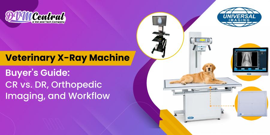
A veterinary X-ray machine is often the fastest way to find out what's going on inside a patient. It helps your team check for fractures, chest and abdominal problems, dental issues, and orthopedic conditions without a lot of guesswork. The right system comes down to who your patients are, how much room you have, who's running the equipment, and how many studies you take on a normal day.
This guide walks through the questions vet teams ask most: which type of system to buy, CR vs. DR, how to get cleaner images, how to position patients, how to keep the workflow moving, and what to do when a radiograph just isn't right.
What Is a Veterinary X-Ray Machine?
A veterinary X-ray machine sends a small, controlled beam of radiation through an animal's body and captures what comes out the other side on a detector. Bone stops most of the beam, so it shows up bright white. Air lets everything through and looks black, while soft tissue like muscle and organs fills in the grays between.
A complete system has four main parts:
- Generator: Produces the beam and sets exposure factors (kVp, mA, and time).
- Tube and collimator: Directs and shapes the beam to the area of interest.
- Table or stand: Supports and positions the patient.
- Detector: Captures and processes the image
- Software: Stores the image.
A vet X-ray machine, animal X-ray machine, and veterinary radiography system are used interchangeably in the market. They all describe the same category of equipment, though configurations vary widely.
What Types of Veterinary X-Ray Machines Are Available?
Most practices choose between a fixed table system, a mobile cart system, or a portable unit. Table systems suit busy hospitals. Mobile carts add flexibility, and portable units work best for field and ambulatory use.
|
System Type |
Best For |
Main Advantage |
Main Limitation |
|
Table-based digital system |
High-volume clinics, specialty hospitals |
Stable, consistent positioning and higher output |
Needs a dedicated room |
|
Mobile cart system |
Small- to mid-sized clinics, multi-room use |
Can move between treatment areas |
Needs careful positioning and floor space |
|
Portable or handheld unit |
Equine, mobile, and farm work |
Goes where the patient is |
Lower output, limited for large body parts |
Choosing an X-Ray Machine for Your Veterinary Practice
The right veterinary X-ray setup depends on the animals you see most often and the size of your largest patients. A system should have enough power for larger dogs while still giving you the control needed for cats and smaller breeds.
Dog X-Ray Machine
A dog X-ray machine needs enough output to produce clear images in larger dogs, especially when imaging the chest or abdomen. Smaller dogs need lower exposure settings, so having good control over the generator makes it easier to adjust for different body sizes.
Cat X-Ray Setup
Cats need more precise exposure settings because of their smaller size. A system with flexible exposure controls lets you adjust the technique without relying on the same settings for every patient.
Generator Power
Generator output affects how quickly an image can be taken. Shorter exposure times can reduce motion blur, which is useful when imaging panting dogs, nervous cats, or patients that have trouble staying still.
- Small animal practice: A mid-range generator may be enough for routine imaging.
- Mixed practice: Higher output gives you more flexibility when imaging larger dogs.
- Equine or large animal practice: Portable units can work well for limb imaging, while body studies generally need more power.
Table Features
A floating tabletop makes it easier to reposition a patient without lifting them. A low table is useful when working with heavier dogs, while a tilting table can make certain views and positioning easier.
Does Species Change the X-Ray Setup?
Yes. Cats, small dogs, large breeds, birds, and reptiles can have different imaging needs. Larger patients may need a bigger detector or more than one exposure. Smaller or exotic patients may benefit from a high-detail detector and suitable restraint.
CR vs. DR: What’s the Difference?
CR and DR are the two main digital radiography options used with veterinary X-ray systems. Both produce digital images, but they work differently.
CR (computed radiography) utilizes a reusable image plate. When the X-ray is taken, the plate is inserted into a reader, which digitizes the image and sends it to a computer. CR might be a good choice for practices that already have compatible X-ray equipment or want to keep the up-front cost low.
DR (digital radiography) uses a digital detector that captures the X-ray image directly. The image appears on the computer within seconds, so there is no separate plate reader to handle.
Here’s a comparison table that helps you pick either a CR or DR X-ray system for your practice.
|
Feature |
CR (Computed Radiography) |
DR (Direct Digital Radiography) |
|
How images are captured |
The cassette is read in a separate scanner. |
The detector sends the image directly to the computer. |
|
Image review time |
Longer, cassette handling needed |
Seconds |
|
Upfront cost |
Lower |
Higher |
|
Workflow |
More steps, more handling |
Fewer steps, fewer retakes |
|
Best fit |
Lower-volume or budget-conscious clinics |
Busy clinics and specialty practices |
Digital X-ray veterinary equipment of either type replaces film and chemicals. It also allows you to adjust brightness and contrast, zoom, measure, annotate, and share photographs with referral specialists.
What Should You Look for in Digital X-Ray Veterinary Equipment?
Look for detector size, image resolution, software usability, generator compatibility, and service support. Fancy features matter less than reliability and ease of use for your team.
Key checkpoints before buying:
- Detector size: Large enough to cover a full canine chest or abdomen in one shot where possible.
- Software: Simple templates, easy measuring tools, and clean PACS or DICOM export.
- Storage and backup: Secure archiving so images are never lost.
- Training: Onboarding for both doctors and technicians.
- Warranty and service: Clear coverage for the tube, generator, and detector.
What Is the Best Veterinary X-Ray Equipment for Orthopedics?
The best veterinary X-ray equipment for orthopedics combines high-detail digital imaging, flexible table movement, and enough detector coverage for joints and long bones. Precise positioning matters more here than in most other imaging.
Orthopedic cases such as cruciate disease, hip dysplasia, elbow dysplasia, fractures, and patellar luxation depend on true, repeatable views. Small positioning errors can hide or mimic disease.
What to prioritize:
- Floating tabletop with locks: Fine adjustments and stable holding.
- High-resolution detector: Clear trabecular and joint detail.
- Measurement tools: Angles and distances for surgical planning.
- Positioning aids: Sandbags, troughs, and foam wedges that keep limbs in place.
- Table size and height: Comfortable handling for large breeds.
An advanced orthopedic X-ray table is designed around these needs, which is useful for hospitals doing frequent hip, stifle, and elbow evaluations or surgical planning.
Veterinary X-Ray Systems Available on DVM Central
DVM Central lists three complete digital radiography options from Universal Imaging. Each one is built to fit a different space, caseload, and budget.
Here are the details.
Complete Mobile Cart-Based X-Ray System
This is a plug-and-play digital system for clinics that need imaging to move between rooms. It includes the generator, panels, computer, display, cart, and software, so there is nothing extra to source. Each component can be customized to your practice needs and budget, and Universal Imaging offers a product specialist consultation about financing and configuration.
Best for: Smaller and mid-sized practices, shared imaging areas, and clinics without a dedicated X-ray room.
Complete Digital X-Ray Table Systems
This table system is designed for clinics with limited space that still see large dogs. The tube column and bucky move horizontally up to 450 mm in either direction, so you can reach the same positions as a float top table without the extra floor space.
- Size: Less than 4 feet wide and about 6 feet tall, with a table large enough for big dogs
- Generator: 5 kW generator with ultrasonic ranging that measures body thickness and suggests exposure settings, plus shortcut keys for 6 groups of exposure parameters
- Detector: 17 by 17 cesium digital receptor, with panels also available in 14 by 17 inch
- Software: iClear acquisition software with bone and soft tissue filters, DICOM PACS mobile viewing, and a built-in positioning guide
- Measurement tools: TPLO, TTA, MMP, and heart measurement (Buchanan's VHS)
Best for: Busy general practices that want a repeatable daily workflow in a small footprint.
Advanced Orthopedics X-Ray Table
This is a four-way float top table with a choice of generator and panel options. It suits hospitals that run a lot of orthopedic work, where fine positioning control matters. The system pairs with most digital imaging setups and is used in general practices, specialty and emergency hospitals, humane societies, zoos, military facilities, and veterinary colleges.
- Generator: 20 kHz high frequency, 30 kW rating, with kVp from 40 to 125 in 1 kV steps
- Tube: 1.0 to 2.0 mm focal spot with 12-foot-high voltage cables
- Table and stand: Enclosed integrated design with a welded base and an animal restraining device
- Collimator: Manual, with a light field selectable at 30, 60, 90, or 120 seconds
Best for: orthopedic and specialty caseloads and hospitals that want a durable table backed by an in-house service team.
Quick Comparison
|
System |
Best For |
Key Strength |
|
Mobile cart system |
Multi-room or space-limited clinics |
Complete, portable, customizable package |
|
Digital table system |
Busy general practice |
Compact table with smart exposure tools and software |
|
Orthopedics table |
Orthopedic and specialty work |
Four-way float top with high-output generator |
How Can You Improve Veterinary X-Ray Image Quality?
Good image quality comes from correct exposure, tight collimation, proper positioning, and reduced patient motion. Equipment matters, but technique matters just as much.
Which Exposure Factors Matter Most?
- kVp: Controls contrast and penetration.
- mA and time (mAs): Controls how much radiation reaches the detector.
- Distance: Keeps geometry consistent from study to study.
Build a technique chart based on patient thickness, and update it whenever equipment changes. Measure the thickest part of the area being imaged, not a guess by eye.
Do You Need a Grid?
For body parts thicker than about 10 centimeters, a grid can reduce scatter and improve contrast. For thin limbs and small patients, it is usually not needed, and using one when unnecessary can raise exposure requirements.
Why Does Collimation Matter?
Tight collimation reduces scatter, improves contrast, and lowers radiation to staff and patient. Always limit the beam to the area of interest, with a small margin.
How Should You Position Animals for Better Radiographs?
Position the patient so the area of interest is straight, centered, and free of rotation. Take at least two views at right angles to each other, since one view can hide a problem.
Standard views by region:
|
Region |
Common Views |
|
Chest |
Right lateral, left lateral, and VD or DV |
|
Abdomen |
Lateral and VD |
|
Hips |
Extended VD and frog leg |
|
Stifle |
Mediolateral and craniocaudal |
|
Elbow |
Mediolateral, flexed lateral, and craniocaudal |
|
Spine |
Lateral and VD, often with sedation |
Is Sedation Necessary During Radiography?
Sedation or anesthesia helps with painful patients, orthopedic views that require exact alignment, and anxious animals. It also reduces the need for manual restraint, which keeps staff hands out of the beam.
How Can You Build a Faster X-Ray Workflow?
A smooth workflow starts with a consistent room setup, a clear technique chart, and a standard order for each type of study. Fewer steps mean fewer retakes and shorter patient stress time.
Practical tips:
- Keep positioning aids within reach of the table.
- Use software templates for common studies.
- Label images at the time of capture, not later.
- Review each image right away before releasing the patient.
- Assign a consistent protocol to each team member.
Digital X-ray systems with DICOM export make referral sharing simple, which speeds up specialist opinions and client communication.
What Are the Most Common Veterinary X-Ray Problems and Fixes?
Most poor images trace back to motion, positioning, exposure, or scatter. Identifying the pattern helps you correct it quickly.
|
Problem |
Likely Cause |
Fix |
|
Blurry image |
Patient motion or long exposure |
Shorten exposure time, use sedation, or use better restraint. |
|
Too dark or too light |
Incorrect mAs or kVp |
Adjust the technique chart, and check patient thickness. |
|
Low contrast, gray look |
Excess scatter or high kVp |
Collimate tighter; consider a grid. |
|
Rotated view |
Poor alignment |
Use positioning aids, and recheck landmarks |
|
Cut off anatomy |
Incorrect centering or detector size |
Center on the region of interest, and use a larger detector. |
|
Artifacts |
Dirty detector or foreign objects |
Clean equipment and remove collars and bandages. |
What Common Mistakes Should Veterinary Teams Avoid?
- Using one technique for every patient size.
- Skipping the second view.
- Ignoring collimation.
- Reviewing images only after the animal has left.
- Buying equipment based on price alone rather than case mix.
- Neglecting staff safety practices such as lead aprons, gloves, and dosimeter badges.
Conclusion
A good veterinary X-ray machine is a combination of the right system type, adequate generator power, a detector that matches your caseload, and consistent technique. DR generally wins on speed and workflow, CR can work for tighter budgets, and orthopedic imaging benefits from precise table movement and positioning tools.
Focus on your real patient mix, train your team on positioning and exposure, and review every image before the patient leaves. Those habits improve diagnosis more than any single feature.
If you are comparing options, DVM Central is a practical source for veterinary X-ray and imaging equipment. You can explore the complete mobile cart-based X-ray system, the complete digital X-ray table systems, and the advanced orthopedic X-ray table to find the setup that fits your practice needs.
Frequently Asked Questions
How much does a veterinary X-ray machine cost?
The price can vary a lot depending on the system, detector, and generator. Portable and CR systems are generally more affordable, while full DR table systems tend to cost more. When comparing options, look beyond the purchase price and factor in installation, training, software, and service costs.
Can one X-ray machine be used for both dogs and cats?
Yes. Most systems designed for general small animal practice can be used for both dogs and cats. What matters is having enough control over exposure settings to adjust for differences in patient size and body thickness.
What is the difference between a dog X-ray machine and a general animal X-ray machine?
In most cases, there is no technical difference. "Dog X-ray machine" is simply a search term often used for equipment intended for small animal practices. The same system can also be used for cats, exotic pets, and other animals when the settings are adjusted to suit the patient.
Is digital X-ray better than film for animals?
Digital X-ray offers several practical advantages over film. Images are available much faster, can be adjusted on the computer, and are easier to store and share. There is also no need for film processing chemicals, and digital systems can make it easier to catch positioning or exposure issues before the patient leaves.
How often does a veterinary X-ray machine need servicing?
Follow the service schedule provided by the manufacturer and have the system inspected and calibrated at the recommended intervals. You should also check your local regulations for any required equipment inspections and radiation safety checks.
Do I need sedation for every X-ray on a dog?
No. Many chest and abdominal X-rays can be taken without sedation when the dog is calm and cooperative. Sedation may be considered for painful or anxious patients and for certain orthopedic, spinal, and dental views where staying still is particularly difficult.
Which is better for a busy clinic, a mobile cart or a table system?
A table system can be a good fit for clinics that take a high number of X-rays because it provides a consistent setup for positioning patients. A mobile cart offers more flexibility and can


DVM Central is the trusted veterinary marketplace connecting suppliers, veterinarians, and pet owners, promoting direct buying for quality animal health products, and simplifying veterinary supply transactions.
%20Blog_Explore%20TicklessUSA%20Chemical-Free%20Tick%20%26%20Flea%20Protection-thumbnail-1790176693.jpg&w=3840&q=75)

%20Blog%20-%20Discover%20Thermotex%20Therapy%20Systems-thumbnail-1789135670.jpg&w=3840&q=75)


